Methodologies · 1000 Springs Project
Sampling & Analysis Protocols
Field sampling techniques, laboratory processing procedures, sequencing protocols, and health & safety guidelines used across the 1000 Springs Project.
Overview diagram
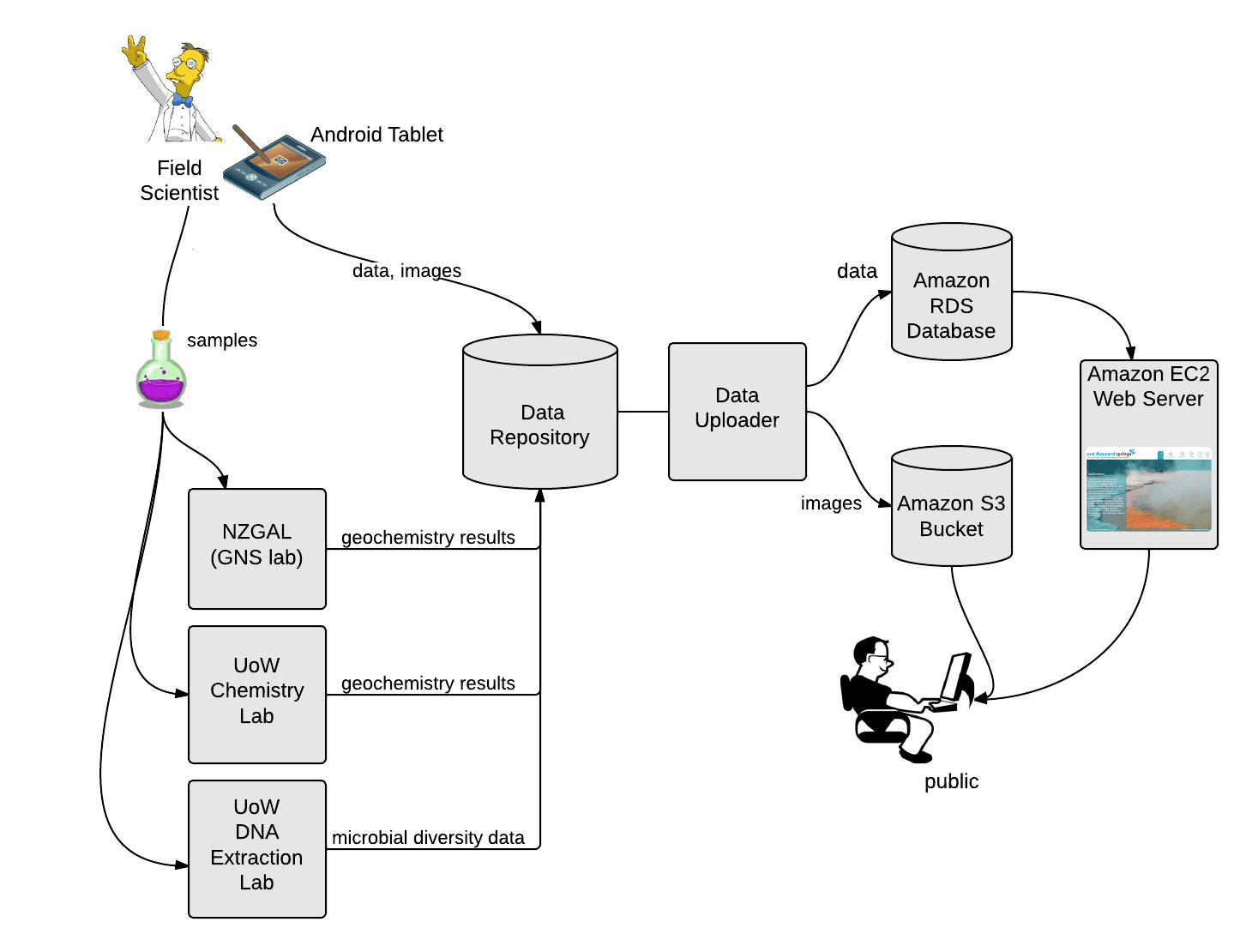
01: Feature Sampling
Sample site selection & water collection
Site selection
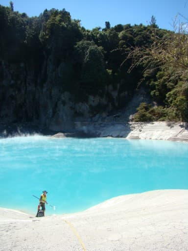
The research targets a broad range of physicochemical conditions in geothermally-influenced springs within the Taupō Volcanic Zone. Water columns are sampled from springs. Sediments, biofilms, mudpots, and heated soils are excluded.
The goal is to collect 1,000 samples, primarily one per individual feature. Repeated sampling occurs where conditions vary significantly, for temporal studies, or for quality control purposes.
Collection technique
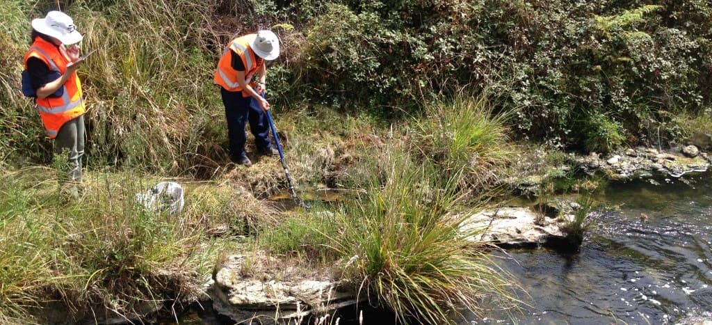
A telescopic sampling pole fitted with sterile containers or custom-built samplers collects water across the full depth of the spring, ensuring homogenous and representative samples.
A field tablet and custom app record physical, chemical, and metadata in the field, eliminating double-handling of field notes and reducing transcription errors. Metadata uploads instantly to the database.
Sample water distribution
Table 1. Volume and container allocation per sample.
| Volume | Container | Purpose |
|---|---|---|
| 2,150 mL | Sterile 2 L polypropylene bottle | Microbial diversity & chemistry analysis |
| ~330 mL | Rubber-necked bottle | H₂S, bicarbonate & chloride analysis |
| 500 mL | Sterile polypropylene bottle | Physical attributes (conductivity, dissolved oxygen) |
| ~60 mL | Syringe | Dissolved gas content |
| ~10 g | Sediment container | Sediment sample (where possible), earmarked for future research |
Field metadata recorded
02: Field & Laboratory Processing
Measurements, filtration & chemistry
Field measurements
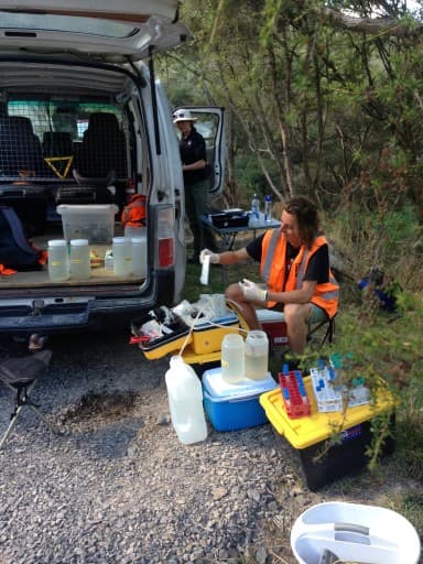
Processing occurs in a 4WD field van configured as a mobile laboratory to minimise temperature changes, dissolved oxygen changes, and microbial activity effects.
Microorganism collection
A battery-operated peristaltic pump pushes up to 2 litres through a 0.2 µm Sterivex column filter (Millipore). After extensive field testing, Sterivex filters demonstrated higher filtration volume capacity than tested alternatives.
Filtrate water is collected in 15 mL and 50 mL tubes for additional chemistry analyses.
All filtrate samples, filters, and soil samples are cooled to 4 °C and transported immediately to the Wairakei laboratory for storage until analysis.
Chemistry analysis locations
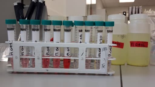
Sample processing summary
Table 2. Container, processing method, storage, and analysis location per parameter.
| Container | Processing | Storage | Parameter | Analytical method | Location |
|---|---|---|---|---|---|
| 2 L bottle | Filtered | −20 °C | Microbial diversity | DNA extraction & sequencing | University of Waikato |
| 500 mL bottle | Raw | n/a | Physical properties | Multiparameter field meter | On site |
| 500 mL bottle | Filtered (0.22 µm) | n/a | Fe²⁺ | UV Spectroscopy | On site |
| 330 mL rubber-sealed | Raw | 4 °C | H₂S | UV Spectroscopy | NZGAL |
| 330 mL rubber-sealed | Raw | 4 °C | HCO₃⁻ | Automated titration | NZGAL |
| 330 mL rubber-sealed | Raw | 4 °C | Cl⁻ | Automated titration | NZGAL |
| 50 mL tube | Filtered | 4 °C | SO₄²⁻ | Ion chromatography | NZGAL |
| 50 mL tube | Filtered | −20 °C | NH₄⁺, PO₄³⁻, NO₂⁻, NO₃⁻ | Flow injection analysis | University of Waikato |
| 50 mL tube | Filtered | 4 °C | Back-up sample | n/a | n/a |
| 15 mL tube | Filtered & acidified | 4 °C | Elements (ICP-MS) | Inductively coupled plasma mass spectrometry | University of Waikato |
| 15 mL tube | Filtered & alkalified | 4 °C | As, Sb | ICP atomic emission spectroscopy | NZGAL |
| 50 mL syringe | Filtered | Room temp. | H₂, CH₄, CO | Gas chromatography | Extremophile Research Group, GNS |
Elements measured via ICP-MS
30 elements quantified by inductively coupled plasma mass spectrometry at the University of Waikato.
03: Sequencing
Microbial diversity assessment
Soil and freshwater can contain up to one billion and ten million cells respectively, making exhaustive identification impractical. The project instead extracts total microbial DNA, sequences a universal gene, and uses bioinformatics pipelines to assign taxonomy at scale using Ion Torrent Next-Generation Sequencing.
DNA Extraction
Summary: Chemical and physical techniques extract, purify, and concentrate DNA from microorganisms. Before cell disruption, sterile skim milk blocks possible binding sites on solids/sediments captured during filtration. Captured cells in the Sterivex filter are disrupted using cetyl trimethylammonium bromide (CTAB). Proteins, lipids, and ribonucleic acids are chemically removed using phenol-chloroform-isoamyl alcohol and chloroform-isoamyl alcohol. DNA is then concentrated via magnetic bead-adsorption.
Cell disruption
Sterivex filters thaw on ice. 1.0 mL of 0.8% w/v skim milk powder solution is added and incubated for 30 min at 65 °C. 0.4 mL CTAB, 0.2 mL phosphate-buffered saline, and 0.1 mL sodium dodecyl sulfate are added and vortexed for 1 minute. The filter is placed in a 50 mL sterile capped tube and incubated at 150 rpm / 65 °C for 30 minutes. After briefly cooling on ice, the aqueous component is pushed through the syringe and collected in 2 mL tubes.
DNA purification
Equal volume (1:1) of chloroform:isoamyl alcohol (24:1) is added to each tube, vortexed for 10 s, and centrifuged at 12,400 rpm for 10 minutes. The aqueous phase (typically top) is transferred to a new tube. 300 µL chloroform:isoamyl (24:1) is added to the aqueous phase, mixed on a Hula mixer for 20 minutes, then centrifuged for 10 minutes at 12,400 rpm. The aqueous phase is collected, avoiding the bottom layer.
DNA concentration
Equal volume of 100% ethanol and 40 µL MoBio beads are added to the aqueous phase. Samples are agitated every 30 s–5 min, then placed on a magnetic stand for 2 minutes. Supernatant is removed and beads are washed with 500 µL of 100% ethanol, cleaning tube walls. All ethanol is removed and beads are resuspended in 30 µL Tris (pH 8.0) by gentle pipetting and brief vortexing.
DNA Amplification & NGS Preparation
Summary: Microbial diversity is determined by sequencing the 16S ribosomal RNA gene (~1,540 nucleotides), a short gene possessed by all bacteria and archaea. Total DNA is extracted and 16S rRNA genes are amplified by PCR modified for Ion Torrent Next-Generation Sequencing. On average, ~70,000 sequences (~270 bp each) are read per sample.
PCR amplification
PCR amplifies the microbial community 16S rRNA genes using universal bacterial/archaeal primers from the Earth Microbiome Project:
5′-GTGCCAGCMGCCGCGGTAA-3′5′-GGACTACVSGGGTATCTAAT-3′The forward primer contains an A-adaptor, a 10–12 nucleotide sample-specific barcode, a 'GAT' barcode adaptor, and the F515 sequencing primer. The reverse primer includes a P1 sequence and R806 sequencing primer.
PCR reaction composition
25 µL total reaction volume. Master mix is pre-treated with ethidium monoazide bromide (EMA) to remove trace DNA contaminants before primer, Taq, and template addition.
Thermocycling
Initial denaturation 94 °C / 3 min → 30 cycles: 94 °C / 45 s · 50 °C / 1 min · 72 °C / 1.5 min → Final extension 72 °C / 10 min
NGS preparation & sequencing
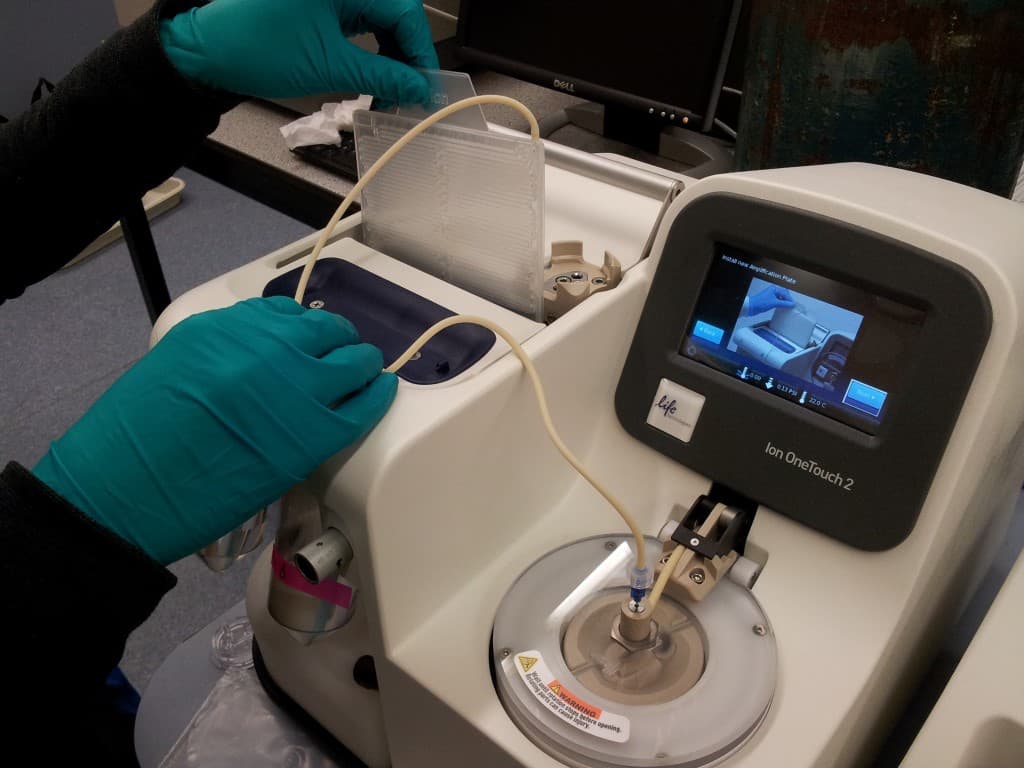
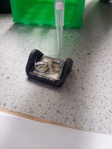
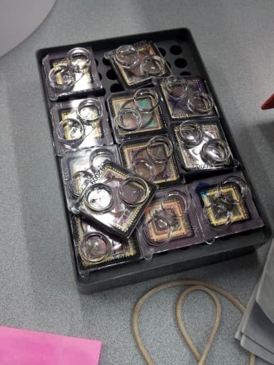
Amplicons from triplicated PCR products are purified using SPRIselect (Beckman Coulter) to remove small nucleotide fragments. Quality and concentration are verified and adjusted to 26 pM via HS Qubit 2.0 (Life Technologies) and 9100 BioAnalyser (Agilent Technologies). Amplicons from all samples are pooled for emulsion PCR. Sequencing is conducted at the Waikato DNA Sequencing Facility using Ion Torrent PGM (Life Technologies) with Ion 318v2 chip and 400 bp chemistry.
Post-sequencing Bioinformatics
Summary: ~70,000 raw reads per sample are processed by customised bioinformatic pipelines. Erroneous reads are removed, tagging sequences are stripped, and reads are screened for known errors and trimmed to 250 bp. Similar sequences are clustered into Operational Taxonomic Units (OTUs) at 97% similarity. OTUs are taxonomically ranked by comparison with microbial databases. For this explorer, OTUs with > 5 reads per sample are presented graphically.
Pipeline details
Raw reads in FASTQ format are processed using a custom pipeline based on Mothur and USEARCH.
04: Health & Safety
Geothermal field safety
Important safety notice
Sampling springs in geothermal environments presents serious danger and should not be attempted by the public. Ground surrounding hot springs can be hot, unstable, and undercut. Springs often emit high concentrations of toxic and/or asphyxiating gases. All project participants have extensive experience moving through and sampling geothermal ecosystems. The project attempts to minimise environmental impact from sampling.
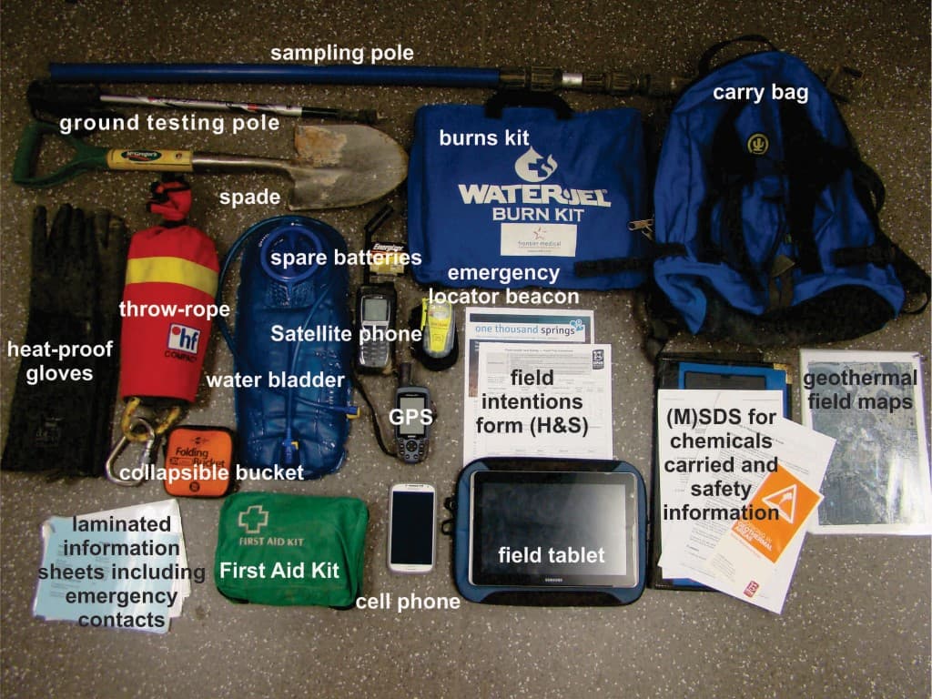
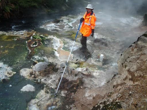
Personal protective equipment & emergency kit
Clothing & footwear
Footwear
Leather boots topped with neoprene puttees (gaiters) are preferred over gumboots; they provide better ankle support and protection in unstable terrain.
Trousers
Locally made Cactus SuperTrousers protect against vegetation and slow water/mud infiltration if accidents occur.
General guidance for visitors
When visiting geothermal environments: do not leave formed tracks, do not approach springs or steaming soils, and always follow posted safety instructions.
References
Literature cited
Edgar, R.C. (2013). UPARSE: Highly accurate OTU sequences from microbial amplicon reads. Nature Methods 10, 996–998.
Rueckert, A. and Morgan, H.W. (2007). Removal of contaminating DNA from polymerase chain reaction using ethidium monoazide. J Microbiol Meth, 68(3), 596–600.
Schloss, P.D., Westcott, S.L., Ryabin, T., Hall, J.R., Hartmann, M., Hollister, E.B., Lesniewski, R.A., Oakley, B.B., Parks, D.H., Robinson, C.J., Sahl, J.W., Stres, B., Thallinger, G.G., Van Horn, D.J. and Weber, C.F. (2009). Introducing mothur: open-source, platform-independent, community-supported software for describing and comparing microbial communities. Appl Environ Microbiol, 75(23), 7537–7541.
Wang Q., Garrity, G.M., Tiedje, J.M. and Cole, J.R. (2007). Naive Bayesian classifier for rapid assignment of rRNA sequences into the new bacterial taxonomy. Appl Environ Microbiol, 73(16), 5261–5267.
Whitman, W.B., Coleman, D.C. and Wiebe, W.J. (1998). Prokaryotes, the unseen majority. Proc Natl Acad Sci USA, 95(12), 6578–6583.
Wilson, A.D. (1960). The micro-determination of ferrous iron in silicate minerals by a volumetric and a colorimetric method. Analyst, 85, 823–827.